


Kontakt aufnehmen
- +49 6221 56 7807
- pankreas@med.uni-heidelberg.de
- EPZ - Europäisches Pankreaszentrum Chirurgische Universitätsklinik, INF 420 69120 Heidelberg
My pancreas is sick
What examinations can I expect?
The doctor will first suspect that something is wrong with the pancreas on the basis of the complaints that the patient describes to him and the physical examination. In order to further confirm this suspicion, and to determine the exact nature of the pancreatic disease, one or more additional examinations, in which the pancreas is imaged, are performed in addition to a blood test.
The following section will briefly describe the various examination methods that are available to determine pancreatic disease.
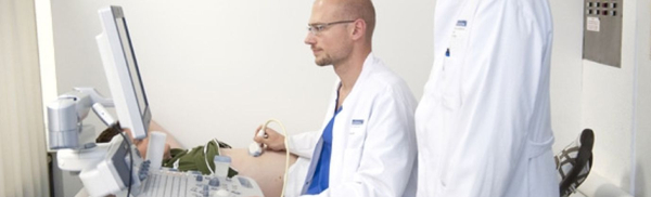
Examination by ultrasound
Ultrasound is the simplest examination to get a picture from the body. Sound waves are sent to the inside of the body through a sensor that the doctor places on the body. These are reflected back by the various organs and registered by the same sensor.
In the process, the sound waves are reflected to different degrees by the individual organs. This creates images on which the abdominal organs, such as the liver, kidney and pancreas, can be identified. According to this image of the organ, it is possible to detect pathological changes.
The examination procedure will be approximately as follows: The examination is performed lying down. Before the transducer is placed on the skin, a gel is applied to improve the contact between the skin and the transducer.
Except for a possible feeling of cold due to the application of the gel, there is no pain or other discomfort associated with this examination. Sometimes it is necessary to hold one's breath for a short time. Air in the bowel may interfere with or make sonography impossible. The ultrasound has no side effects.
Examination by X-rays
This is probably the most frequently performed examination for pancreatic diseases. The computer tomogram works with X-rays. By taking a large number of cross-sectional images through the body, it is possible to get a precise impression of the structure of the pancreas and surrounding organs.
The examination will proceed approximately as follows: About half an hour before the examination, the patient must drink a special liquid so that the stomach and intestines can later be easily distinguished from the other organs in the images. In the X-ray room, the patient lies on a couch that can be moved automatically. He receives instructions and information from the control room via an intercom system. The couch with the patient is guided through a tube about 50 cm long, and the cross-sectional images are taken during this process.
During the second half of the examination, a contrast medium is injected into the arm vein so that the vessels and abdominal organs can be better visualized. This section is occasionally omitted. The whole procedure takes about half an hour.
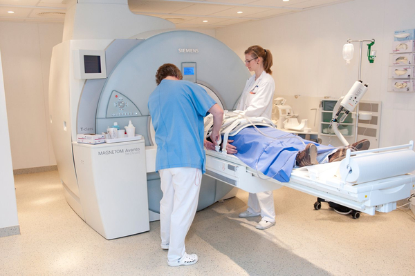
Examination with the help of magnetic fields
The magnetic resonance examination is a similar examination to the computer tomography. Here, too, cross-sectional images of the body are made. The examination does not use X-rays, but works with the help of changing magnetic fields.
For this purpose, the patient must lie down in a tube and try to remain still as much as possible during the examination. Depending on the question, the ductal system, blood vessels and glandular tissue of the pancreas can be particularly visualized here.Since magnetic fields are used here, the patient must indicate before the examination whether he or she is wearing metal prostheses or metal fragments from the war in the body. Depending on the technique, magnetic resonance imaging generates noise pollution.
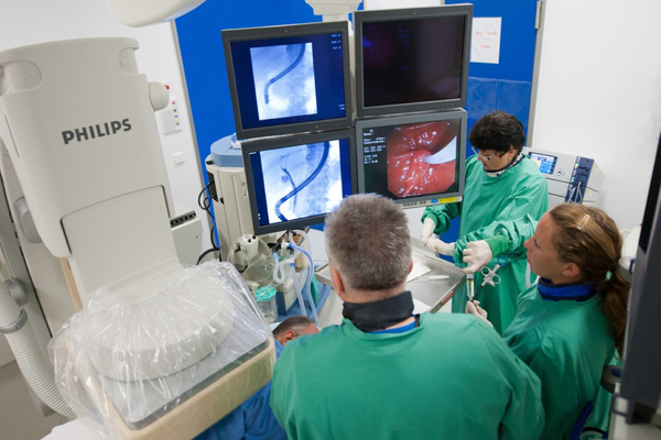
Examination by means of gastroscopy
ERCP is used to obtain a precise impression of the bile ducts and pancreatic ducts. This is a very important complementary examination to the other imaging examinations.
In this procedure, a thin catheter is inserted via a special gastroscope into the excretory duct of the pancreas or bile ducts (papilla Vateri), which is located in the duodenum. A contrast medium is then injected through this catheter and the ducts thus visualized are X-rayed. If necessary, a drain can be inserted during the examination to improve the drainage of secretions into a blocked duct, stones can be removed, or the excretory duct can be widened with an electric knife.
Ultrasound examination from the inside
Endosonography is a method in which the esophagus, stomach and duodenum are examined with a probe. It involves the use of a flexible examination tube, also called an endoscope, which is equipped with optics and an ultrasound probe at the tip. This endoscope is inserted into the body through the mouth, allowing for precise and effective diagnosis. With the help of this advanced technology, diseases and injuries in the affected areas can be detected quickly and reliably.
